STUDENT CENTER
Get Your Degree!
Find schools and get information on the program that’s right for you.
Powered by Campus Explorer
Welcome to Radiology
Medical Imaging is a cornerstone of modern healthcare, allowing clinicians to visualize the internal structures and functions of the body without the need for invasive procedures. By using technologies such as X‑ray, computed tomography (CT), magnetic resonance imaging (MRI), ultrasound (US), nuclear medicine (NM), and positron emission tomography (PET), medical imaging modalities help detect disease, guide treatment, and monitor patient progress. Students and future students can find resources here. Some examples: radiology career guidance, radiology encyclopedia, radiology term paper ideas, radiology licensure and more. You will also discover respiratory therapy resources and other allied health information.
X-ray Schools
X‑ray schools, often part of accredited radiologic technology programs, provide students with the foundational knowledge and clinical training needed to become radiographers. These programs typically include coursework in anatomy, radiation physics, patient care, image production, and radiation safety, along with hands‑on clinical rotations in hospitals or imaging centers. Most programs award an associate degree, though some institutions offer bachelor’s pathways for advanced study. Graduates are eligible to sit for the ARRT certification exam, which is required for licensure in most states and serves as a key credential for entering the profession.
X-ray Jobs
X‑ray jobs span a wide range of clinical environments, including hospitals, outpatient imaging centers, urgent care facilities, orthopedic practices, and mobile imaging services. Radiographers perform diagnostic imaging exams, ensure patient safety, maintain equipment, and collaborate with radiologists and other healthcare professionals. Career paths may expand into specialized modalities such as CT, MRI, mammography, or interventional radiology, as well as leadership roles in education, management, or quality assurance. Demand for radiologic technologists remains strong due to ongoing healthcare needs and the essential role imaging plays in modern diagnosis and treatment.
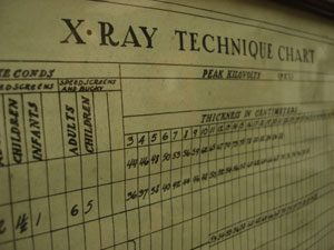
X-ray Techniques
X‑ray imaging relies on a variety of techniques designed to optimize diagnostic quality while minimizing radiation exposure. Common methods include standard projection radiography, fluoroscopy for real‑time imaging, and specialized approaches such as contrast studies, mobile radiography, and digital radiography. Proper patient positioning, exposure selection, collimation, and use of protective shielding are essential components of technique. Radiographers must understand how factors like kilovoltage, milliamperage, distance, and motion affect image clarity and diagnostic value, ensuring that each exam meets clinical standards.
Radiology Categories
Allied Health Clinical Preparation and Readiness College Overview and Campus Basics College Selection and Decision Making Computed Tomography Core Radiology Coursework CT CEU Topics CT Paper Ideas DEXA Exams Dictionary General CEU Topics General Paper Ideas Housing, Dining, and Campus Services Imaging Modalities and Techniques Interventional Paper Ideas Interventional Radiology LPN Mammography MRI CEU Topics MRI Paper Ideas Nuclear Medicine Nuclear Medicine Paper Ideas PET Professional Growth and Clinical Confidence Radiation Physicist Radiation Therapy Radiography Radiology Radiology Encyclopedia Radiology History Radiology Pacs Radiology Physician Radiology Textbooks and Study Resources Rad Tech State License Recommendation Letters and Support Materials Respiratory Therapist Respiratory Therapy RN Safety, Compliance, and Clinical Protocols Social Life and Belonging Student Life and Campus Resources Ultrasound Ultrasound CEU Topics Ultrasound Paper Ideas X ray CEU Topics
Radiology Sources
- Radiological Society of North America (RSNA). RadiologyInfo.org – Patient Information on Medical Imaging Modalities. RSNA & ACR.
- American College of Radiology (ACR). Practice Parameters and Technical Standards for Medical Imaging.
- American Registry of Radiologic Technologists (ARRT). Radiography, CT, MRI, Sonography, and Nuclear Medicine Certification Content Specifications.
- National Institutes of Health (NIH). MedlinePlus – Diagnostic Imaging.
- Society of Nuclear Medicine and Molecular Imaging (SNMMI). Nuclear Medicine & Molecular Imaging Overview.
- American Society for Radiation Oncology (ASTRO). Radiation Therapy Basics.
- U.S. Food & Drug Administration (FDA). Mammography Quality Standards Act (MQSA) – Breast Imaging Information.
- International Society for Clinical Densitometry (ISCD). Bone Densitometry Guidelines and Position Statements.
- Healthcare Information and Management Systems Society (HIMSS).
Medical Imaging Topic Areas
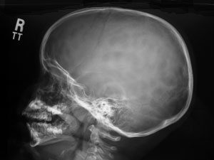
Radiography
Radiography introduces students to the foundational principles of X‑ray imaging, supported by resources such as radiography textbooks, practice guidelines, and equipment information. Learners explore how X‑rays interact with tissue, emphasizing positioning, radiation safety, and image‑quality optimization.
Cat Scan
Computed tomography uses rotating X‑ray beams and advanced reconstruction algorithms to create detailed cross‑sectional images. Students can deepen their knowledge through CT textbooks, protocol guides, and equipment overviews.
Magnetic Resonance
MRI provides exceptional soft‑tissue contrast using magnetic fields and radiofrequency pulses. Learners can explore MRI textbooks, clinical guidelines, and equipment resources to understand physics, safety, and clinical applications.
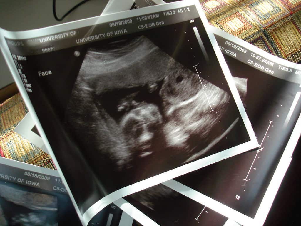
Ultrasound
Ultrasound uses high‑frequency sound waves for real‑time imaging of soft tissues and blood flow. Students benefit from ultrasound textbooks, protocol resources, and equipment guides.
Nuclear Medicine
Nuclear medicine evaluates organ function and metabolism using radiopharmaceuticals. Learners can explore nuclear medicine textbooks, guidelines, and equipment information.
PET Scan
PET imaging detects metabolic activity using positron‑emitting tracers. Students can access PET textbooks, protocols, and equipment resources to understand its role in oncology, neurology, and cardiology.
Mammography
Mammography uses low‑dose X‑rays to detect breast abnormalities. Students can explore mammography textbooks, guidelines, and equipment details.
Interventional
Interventional radiology uses imaging guidance to perform minimally invasive procedures. Learners can review IR textbooks, protocols, and equipment resources.
Radiation Therapy
Radiation therapy uses targeted ionizing radiation to treat cancer and other conditions. Students can explore radiation therapy textbooks, guidelines, and equipment information.
Bone Densitometry
Bone densitometry, commonly performed with DEXA, measures bone mineral density for osteoporosis assessment. Learners can access DEXA textbooks, guidelines, and equipment resources.
Radiology PACS
PACS systems store, manage, and distribute digital medical images. Students can explore PACS textbooks, architecture resources, and workflow guides.
Radiologist
Radiologists interpret medical images and guide patient management. Students can explore radiology physician textbooks, guidelines, and professional resources.